60 Animal Cell Under Transmission Electron Microscope
The image is magnified and focused onto an imaging device such as a fluorescent screen or a. Some variation of this microscope can also penetrate down to the subatomic particles like electrons.
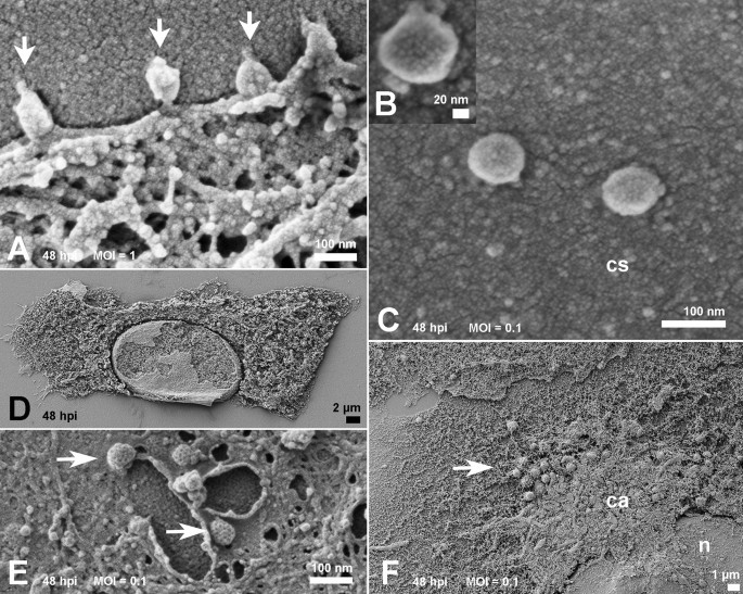
Ultrastructural Analysis Of Sars Cov 2 Interactions With The Host Cell Via High Resolution Scanning Electron Microscopy Scientific Reports
Spirogyra Under Microscope 10x.
Animal cell under transmission electron microscope. Methods In Cell Biology. Methods In Cell Biology. Blood opens an overview page on the different blood cells.
The cell wall nucleus vacuoles mitochondria endoplasmic reticulum Golgi apparatus and ribosomes are easily visible in this transmission electron micrograph. You see that many features are in common. Magnification 50 000x Transmission Electron Micrograph Tem.
Monster Designs Animal Cell Under An Electron Microscope. Microscope Cell Images Animal Cells All Living Things Are Made Up. Under the terms of the Creative Commons Attribution License which permits unrestricted use distribution and reproduction in any medium provided the.
Charles Daghlian Dartmouth Electron Microscope Facility via Wikimedia Commons. However no obvious structural damage was apparent and several repeated scans gave the same images. Commencing circa 1950 the development of the transmission electron microscope revolutionized microscopy progressively bringing it.
This would allow the cell to be viewed in a lot more detail than what it was with a light microscope however the image is only ever produced in shades grey. Fast Transmission Electron Microscope TEM. The microscope can not only distinguish between individual atoms but even see them when they were about only 04 angstroms apart half the length of a chemical bond.
Exercise 3 Cells Organelles And Inclusions. Liquid cell electron microscopy e. The plant cell as more rigid and stiff walls.
The diagram below shows the general structure of an animal cell as seen under an electron microscope. The nature of the image depends on the type of light or electron microscope used and on the way in which the cell or tissue has been prepared for observation. Thats the major difference between plant and animal cells under microscope.
Human and animal mitochondria. You know Animal cell structure contains only 11 parts out of the 13 parts you saw in the plant cell diagram because Chloroplast and Cell Wall are available only in a plant cell. Usb Microscope Software Windows 10.
Isolated mitochondria as well as cell suspensions to be completed in. However the transmission electron microscope uses a high voltage beam which passes through a thin sample showing the internal structure of the cell. These are both specific types of.
Table D leads to images of electron microscopes or protocols for tissue preparation. Electron microscope The images from the transmission electron microscope show a razor-thin layer just two atoms thick of two atoms bonded together. The development of electron microscopes has greatly extended the ability to resolve sub-cellular particles and it has provided new information on the organization of plant and animal tissues.
Trachea epithelium showing ciliated cells cells with hair-like projections. The Great Story Iii An Animal Cell A Eukaryote Before The. Transmission Electron Microscope Tem Micrograph Showing Several.
Below the basic structure is shown in the same animal cell on the left viewed with the light microscope and on the right with the transmission electron. Transmission Electron Microscope TEM Zoology for IAS IFoS and other competitive exams. Here is an electron micrograph of an animal cell with the labels superimposed.
In this lab you observe typical undifferentiated plant cells parenchymaYou should have note the characteristics that plant cells share with all other eukaryotic cells the nucleus and membrane-bound organelles and also note the characters where plant cells differ from animal cells large central vacuole plastids and cell wall. Animal cells have a basic structure. All you need for this is a microscope with a basic transmitted light source and enough magnification to resolve individual yeast cells.
Transmission and some scanning electron microscopic images oforgans. Head and mouth of a. In transmission electron microscope TEM a beam of electrons is transmitted through the section of a specimen and an image is formed by the interaction of the electrons transmitted through the specimen.
The animal cell is more fluid or elastic or malleable in structure. What can be seen under a electron microscope. Under the intense radiation of the electron microscope 011 electron per Å 2 the question of viability of cells naturally arises because the amount of radiation absorbed during highmagnification imaging is sufficient to cause cell death.
The transmission electron microscope.
20+ Animal And Plant Cell Diagram Labeled
Plant And Animal Cells Diagram Quiz Biological Science Picture Unlabeled Blank Animal Cell Diagram To Label Animal Cell To Label 7th Grade Cell Worksheets Globalexotica Net. A cell wall - normally composed of cellulose- on the outside of the cell.

Cells Lessons Blendspace
Plant Cell Diagram Black And White.

Animal and plant cell diagram labeled. Unlabeled animal and plant cell diagram. Listed below are the Cell Organelles of an animal cell along with their functions. An improved method to screen Fc function of immunoglobulin products was developed using CMV kodecytes10 and FSL antigen constructs have been printed onto silica to create a convenient array for antibody identification4 Measles virions MV were fluorescently labeled to monitor binding to DC-SIGN expressing CHO cells using flow cytometry7 Carbohydrate FSL Constructs11 have been.
Square or rectangular in shape. Allows materials in and out. Present and lies in the centre of the cell.
A bacteria diagram clearly helps us to benefit extra about this unmarried cell organisms that have neither membrane-bounded nucleolus or organelles. 7 rows Animal cells usually have an irregular shape and plant cells usually have a regular shape. The typical characteristics that define the plant cell include cellulose hemicellulose and pectin plastids which play a major role in photosynthesis and storage of starch large vacuoles responsible for regulating the cell.
This is rigid so it gives the plant cell its shape. Have chloroplasts and use photosynthesis to produce food have cell wall made of cellulose A plant cell has plasmodesmata - microscopic channels which traverse the cell walls of the cells one very large vacuole in the center are rectangular in shape Animal Cells. While animal cells may have many tiny vacuoles a plant cell usually has a single large vacuole which serves as a storage tank for food water waste products and other materials.
The vacuole has an important structural function as well. Irregular or round in shape. There are various organelles present within the cell and are classified into three categories based on the presence or absence of membrane.
Also Read Different between Plant Cell and Animal Cell. Animal Cell Nucleus Plant Cell Cytoplasm Cell membrane Mitochondria Vacuole Chloroplast Cell Wall. Plant Cell Diagram Animal Cell Diagram Featured in this printable worksheet are the diagrams of the plant and animal cells with parts labeled vividly.
Learn the cell structures and how to identify and label themLike. What Is An Animal Cell Animal Cell Model Diagram Project Parts Structure Labeled Coloring and Plant Cell Organelles Cake. The structure labeled e is called cpu of the cell where the genetic material resides.
Which of the following organelles does an animal cell NOT have. The Cell Organelles are membrane-bound present within the cells. Which of the following pair structures do not have similar functions.
Present and lies on one side of the cell. This may be useful as a printable poster. Blank Animal Cell Diagram To Label Human Body Anatomy Animal Cell Clipart Labeled.
It is made of several layers of parallel fibres and the gaps are filled in with jelly-like substances eg. Assembling cells from ready-made components in form of stickers. Nucleolus A round structure in the nucleus that makes ribosomes.
Present but are very rare. Well-Labelled Diagram of Animal Cell. PLANT AND ANIMAL CELLPLANT AND ANIMAL CELLS SSS Organelle Function Cell Membrane A double layer that supports and protects the cell.
Biology Multiple Choice Quizzes Plant Cell And Animal Cell Diagram Quiz Unlabeled animal cell diagram. The cell membrane is a double-layered membrane made up of phospholipids that surrounds the entire cell. Vacuole Stores food and water.
This enhanced visual instructional tool assists in grasping and retaining the names of the cell parts like mitochondrion vacuole. In addition to the chloroplast the mitochondrion and the nucleus are cut through revealing their internal structures.
