The image is magnified and focused onto an imaging device such as a fluorescent screen or a. Some variation of this microscope can also penetrate down to the subatomic particles like electrons.
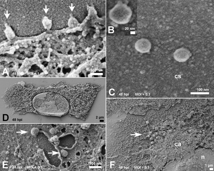
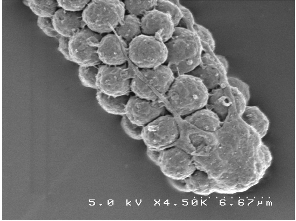
Ultrastructural Analysis Of Sars Cov 2 Interactions With The Host Cell Via High Resolution Scanning Electron Microscopy Scientific Reports
Spirogyra Under Microscope 10x.
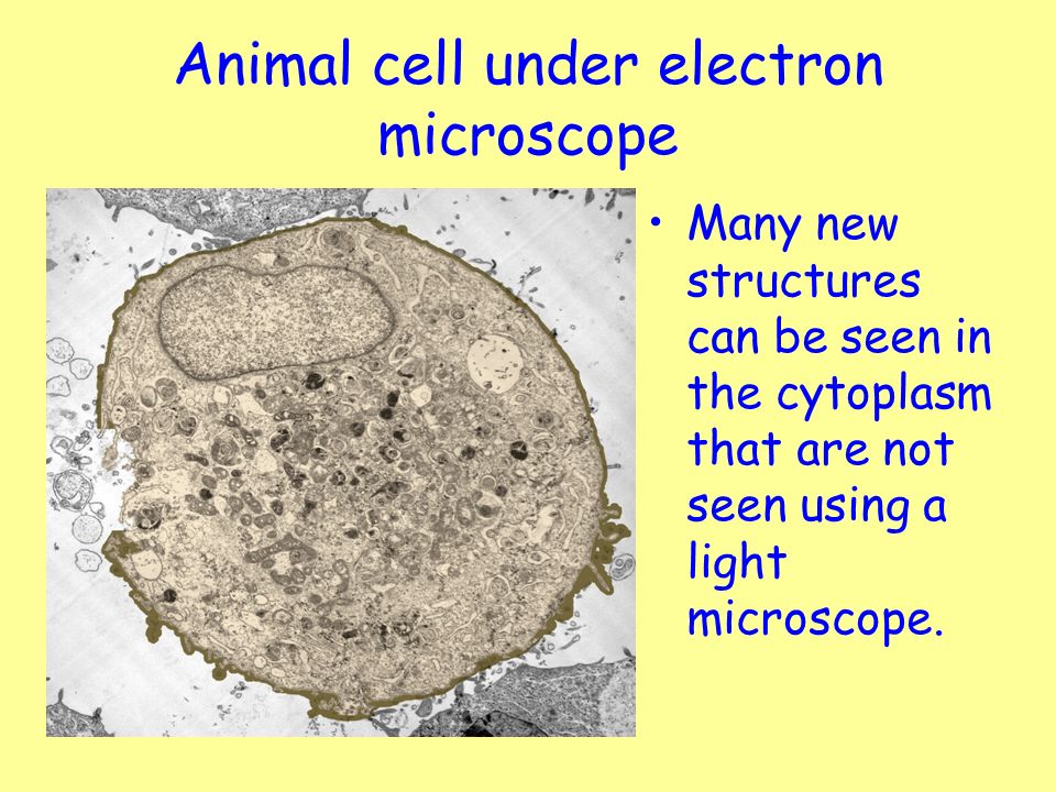
Animal cell under transmission electron microscope. Methods In Cell Biology. Methods In Cell Biology. Blood opens an overview page on the different blood cells.
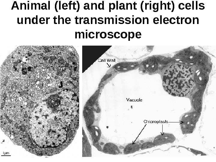
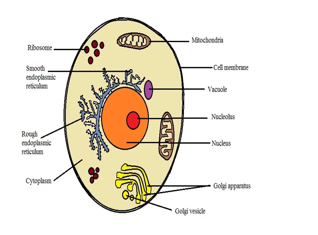
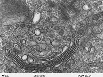
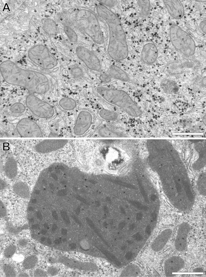
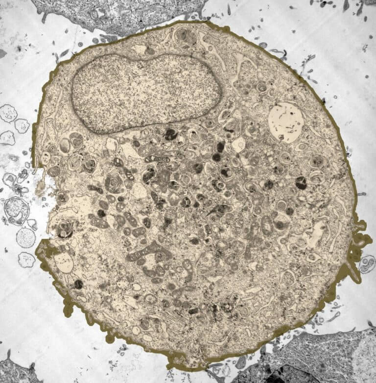
The cell wall nucleus vacuoles mitochondria endoplasmic reticulum Golgi apparatus and ribosomes are easily visible in this transmission electron micrograph. You see that many features are in common. Magnification 50 000x Transmission Electron Micrograph Tem.
Monster Designs Animal Cell Under An Electron Microscope. Microscope Cell Images Animal Cells All Living Things Are Made Up. Under the terms of the Creative Commons Attribution License which permits unrestricted use distribution and reproduction in any medium provided the.
Charles Daghlian Dartmouth Electron Microscope Facility via Wikimedia Commons. However no obvious structural damage was apparent and several repeated scans gave the same images. Commencing circa 1950 the development of the transmission electron microscope revolutionized microscopy progressively bringing it.
This would allow the cell to be viewed in a lot more detail than what it was with a light microscope however the image is only ever produced in shades grey. Fast Transmission Electron Microscope TEM. The microscope can not only distinguish between individual atoms but even see them when they were about only 04 angstroms apart half the length of a chemical bond.
Exercise 3 Cells Organelles And Inclusions. Liquid cell electron microscopy e. The plant cell as more rigid and stiff walls.
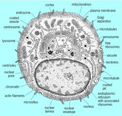
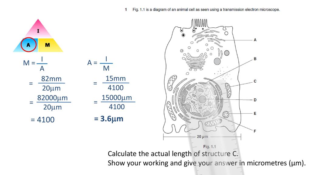
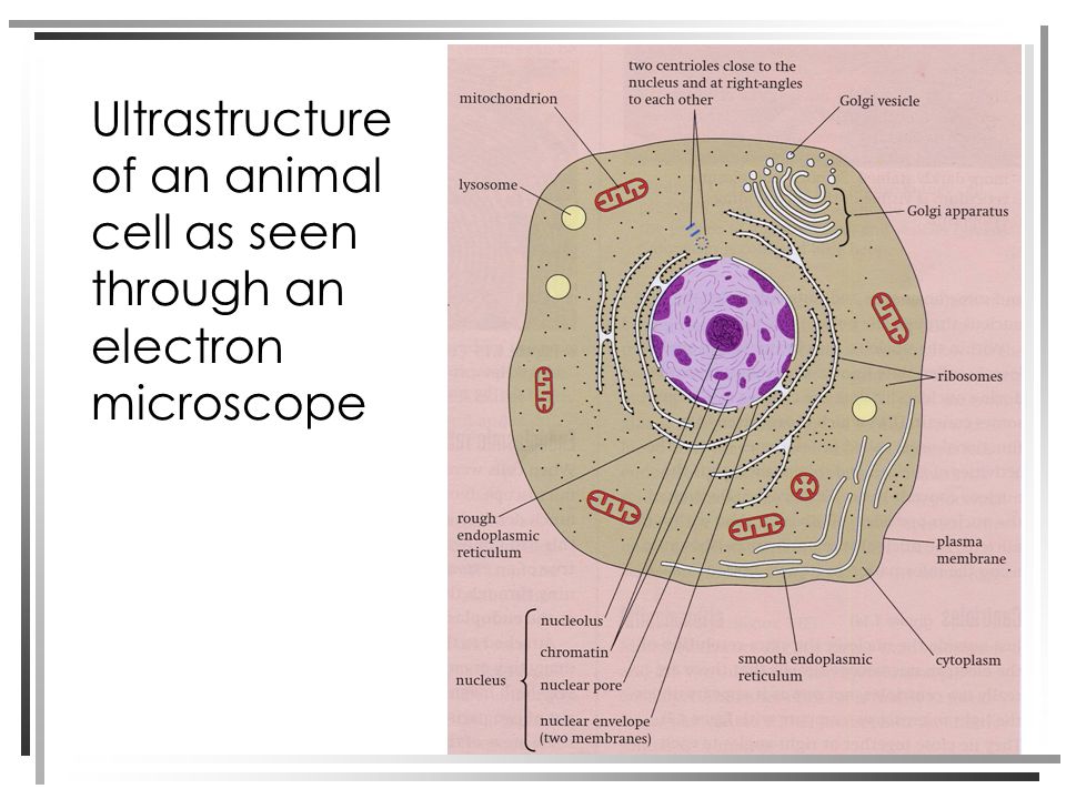
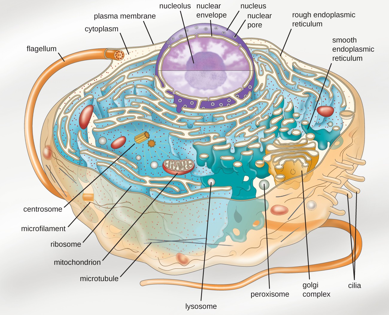
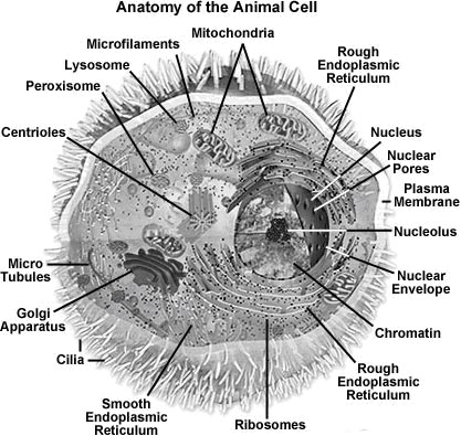
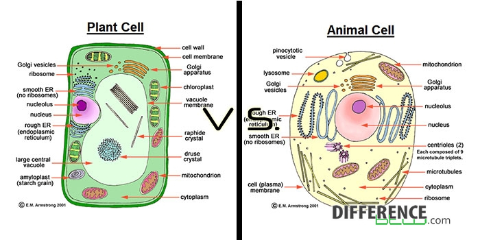
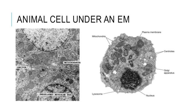
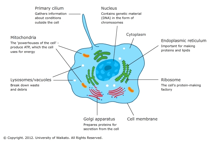
The diagram below shows the general structure of an animal cell as seen under an electron microscope. The nature of the image depends on the type of light or electron microscope used and on the way in which the cell or tissue has been prepared for observation. Thats the major difference between plant and animal cells under microscope.
Human and animal mitochondria. You know Animal cell structure contains only 11 parts out of the 13 parts you saw in the plant cell diagram because Chloroplast and Cell Wall are available only in a plant cell. Usb Microscope Software Windows 10.
Isolated mitochondria as well as cell suspensions to be completed in. However the transmission electron microscope uses a high voltage beam which passes through a thin sample showing the internal structure of the cell. These are both specific types of.
Table D leads to images of electron microscopes or protocols for tissue preparation. Electron microscope The images from the transmission electron microscope show a razor-thin layer just two atoms thick of two atoms bonded together. The development of electron microscopes has greatly extended the ability to resolve sub-cellular particles and it has provided new information on the organization of plant and animal tissues.
Trachea epithelium showing ciliated cells cells with hair-like projections. The Great Story Iii An Animal Cell A Eukaryote Before The. Transmission Electron Microscope Tem Micrograph Showing Several.
Below the basic structure is shown in the same animal cell on the left viewed with the light microscope and on the right with the transmission electron. Transmission Electron Microscope TEM Zoology for IAS IFoS and other competitive exams. Here is an electron micrograph of an animal cell with the labels superimposed.
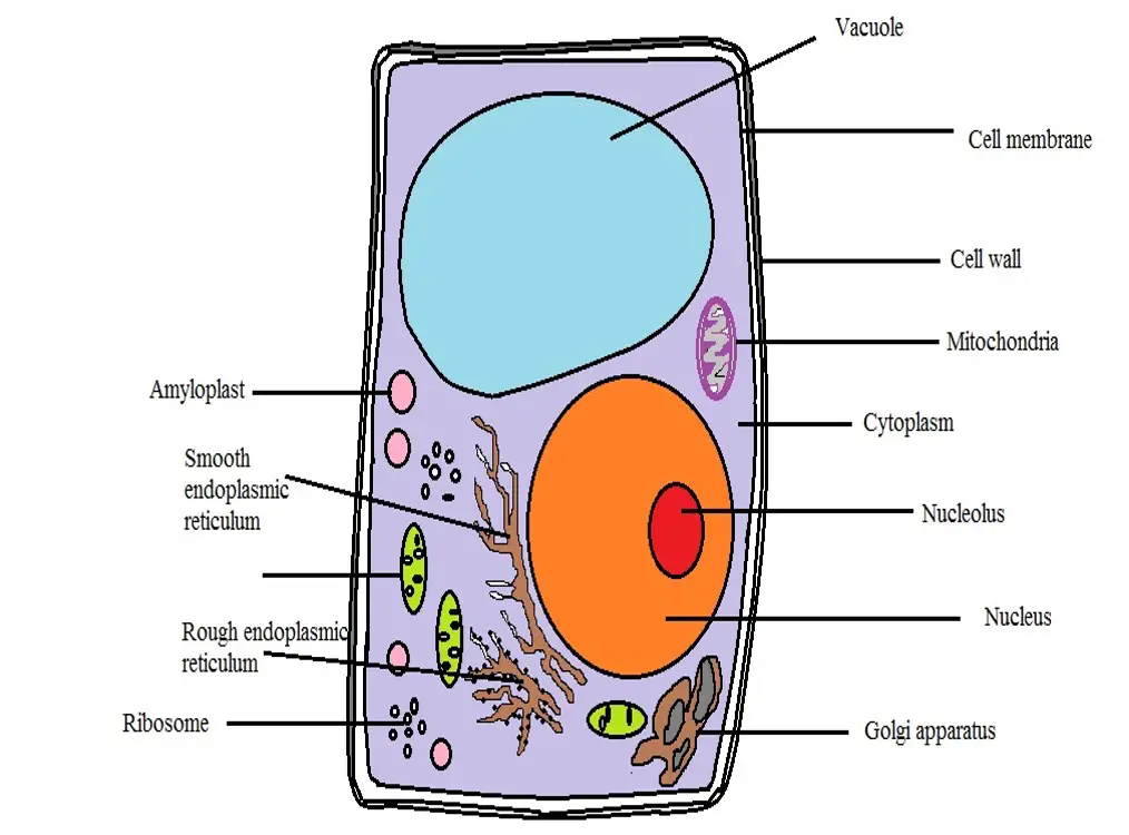
In this lab you observe typical undifferentiated plant cells parenchymaYou should have note the characteristics that plant cells share with all other eukaryotic cells the nucleus and membrane-bound organelles and also note the characters where plant cells differ from animal cells large central vacuole plastids and cell wall. Animal cells have a basic structure. All you need for this is a microscope with a basic transmitted light source and enough magnification to resolve individual yeast cells.
Transmission and some scanning electron microscopic images oforgans. Head and mouth of a. In transmission electron microscope TEM a beam of electrons is transmitted through the section of a specimen and an image is formed by the interaction of the electrons transmitted through the specimen.
The animal cell is more fluid or elastic or malleable in structure. What can be seen under a electron microscope. Under the intense radiation of the electron microscope 011 electron per Å 2 the question of viability of cells naturally arises because the amount of radiation absorbed during highmagnification imaging is sufficient to cause cell death.
The transmission electron microscope.

Net Formation In Peritoneal Colon Cancer Metastasis In Mice A Download Scientific Diagram
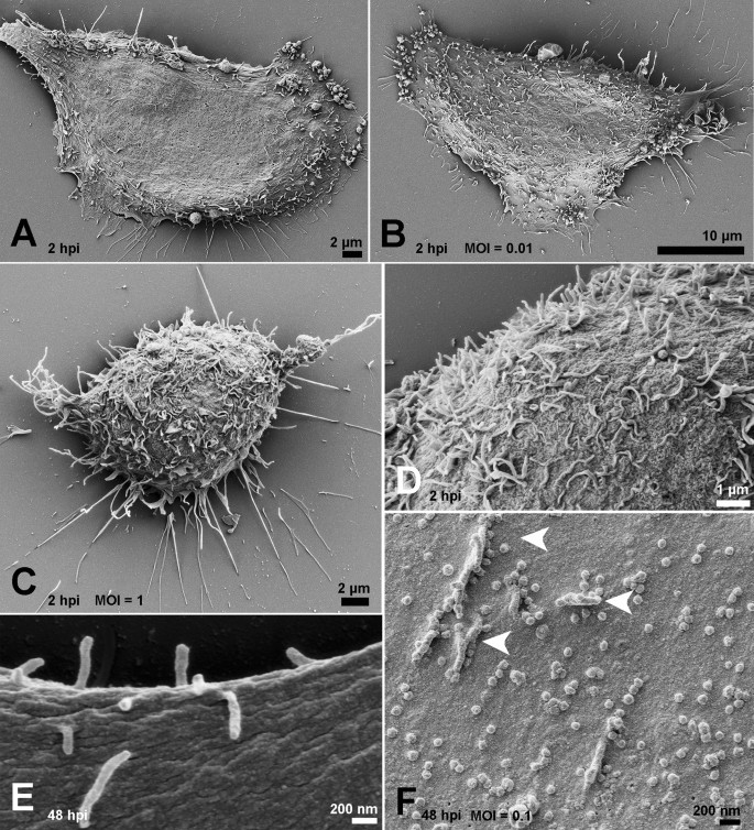
Ultrastructural Analysis Of Sars Cov 2 Interactions With The Host Cell Via High Resolution Scanning Electron Microscopy Scientific Reports
What Does An Animal Cell Look Like Under An Electron Microscope Quora
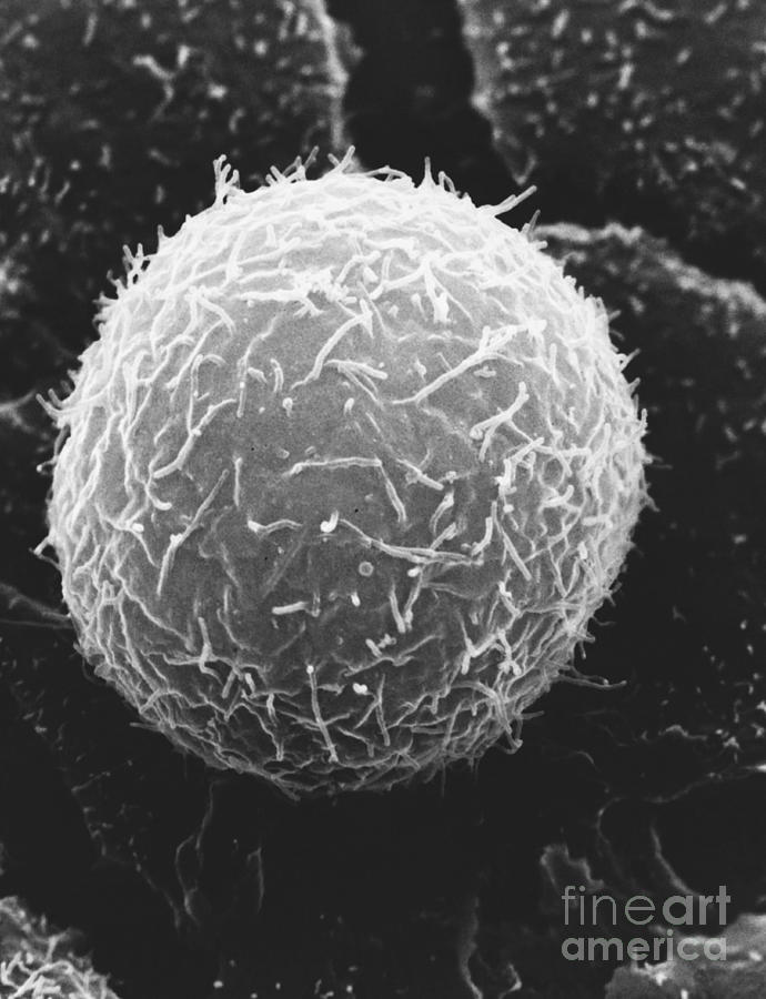
Typical Animal Cell Sem Photograph By David M Phillips
Cell Structure Article About Cell Structure By The Free Dictionary
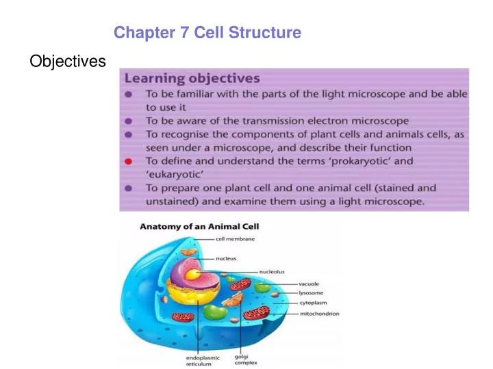
Microscopy And Magnification Ppt Download
Transmission Electron Micrograph High Res Stock Images Shutterstock
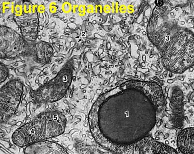
1
Frontiers Large Volume Electron Microscopy And Neural Microcircuit Analysis Frontiers In Neural Circuits
What Is Electron Microscopy Umass Medical School
Puzzle Of The Day What Is The Link
![]()
Lysosome High Resolution Stock Photography And Images Alamy
Structure Of Plant And Animal Cells Under An Electron Microscope Ppt Video Online Download
Animal Cell High Resolution Stock Photography And Images Alamy
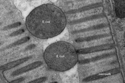
Scientists Offer First Definitive Proof Of Bacteria Feeding Behavior In Green Algae
![]()
Transmission Electron Microscope Tem Micrograph Of A Gonadotropic Stock Photo Picture And Royalty Free Image Image 97108391

Electron Microscope An Overview Sciencedirect Topics
Cellular Organization


Virus Detection By Transmission Electron Microscopy Still Useful For Diagnosis And A Plus For Biosafety Roingeard 2019 Reviews In Medical Virology Wiley Online Library
Low Voltage Electron Microscope Lvem 25 Delong Instruments


Scanning Electron Microscope Sem Images Of The Three Cell Lines Of Download Scientific Diagram
1
Methods In Cell Biology
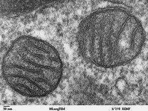
Mitochondrion Wikipedia
![]()
Types Of Microscopes 1 Compound Light Microscope Ppt Download
![]()
Lysosome High Resolution Stock Photography And Images Alamy
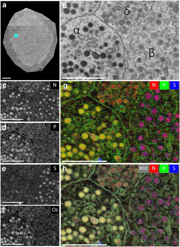
Multi Color Electron Microscopy By Element Guided Identification Of Cells Organelles And Molecules Scientific Reports
Cellular Organization

![]()
Transmission Electron Microscopy Tem Service Creative Bioarray

Chapter 3 Cell Structure And Function Concepts Of Biology

Electron Microscopes Cell Structure Edexcel Gcse Combined Science Revision Edexcel Bbc Bitesize

Eukaryotic Cell Animal Cell Animal Biology Cell En Eukaryotic Membrane Nucleus Science Template Tp Glogster Edu Interactive Multimedia Posters
Electron Micrographs
32 Label The Transmission Electron Microscope Image Of A Chloroplast Below Labels Database 2020
Sample Preparation In Tem
Electron Micrographs
![]()
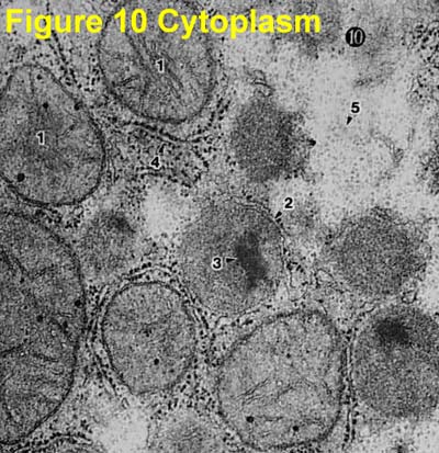
Cytoplasm Cell Organelles Tem Stock Image Image Of Reticulum Lysosomes 101296169

Characterizing Viral Infection By Electron Microscopy The American Journal Of Pathology
Cell Organelles Science Learning Hub

Mitochondria Creationwiki The Encyclopedia Of Creation Science
Animal Cell Tem Stock Image C013 1439 Science Photo Library


Magnification 50 000x Middot Transmission Electron Micrograph Tem Showing Golgi Apparatus Of Chlamydomonas Alg Cell Organelles Macro And Micro Animal Cell
What Are The Differences Between A Plant Cell And An Animal Cell
Morphological Aspects Of Apoptosis
What Is A Diagram Of A Plant And Animal Cell Under An Electron Microscope Quora
Rem Tem Images Of Plant And Animal Cells Hampden Academy Biology 2012

Ribosome Associated Vesicles A Dynamic Subcompartment Of The Endoplasmic Reticulum In Secretory Cells Science Advances

12 Differences Between Scanning Electron Microscope And Transmission Electron Microscope Sem Vs Tem

Scanning Electron Microscopy Of Cells And Tissues Under Fully Hydrated Conditions Pnas
Ultrastructure

Cil 7724 Rattus Epithelial Cell Epithelial Cell Of Thymus Cil Dataset
What Cell Organelles Can Be Seen Under The Electron Microscope But Not With The Light Microscope And Their Functions In The Cell Quora
Electron Micrographs
Cell Structure Learning Intention Ppt Video Online Download
Transmission Electron Micrograph Of Animal Cell Stock Image G450 0051 Science Photo Library

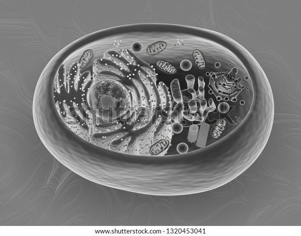
Animal Cell Scanning Electron Microscope Imitation Stock Illustration 1320453041

Animal Cell Definition Structure Parts Functions And Diagram
Introduction To Electron Microscopy Advanced Microscopy Imaging Facilities The University Of Utah

Transmission Electron Microscopy An Overview Sciencedirect Topics

Let S Talk About The Cell Part One Biochemunclassified
Https Encrypted Tbn0 Gstatic Com Images Q Tbn And9gcqdfvm3em56wxups9pla5fodpbggh2d7b2qxnocxw Xur0pfr2m Usqp Cau

Transmission Electron Microscopy Studies Of Cellular Responses To Entry Of Virions One Kind Of Natural Nanobiomaterial

Difference Between Plant And Animal Cells Cells As The Basic Units Of Life Siyavula

Virus Detection By Transmission Electron Microscopy Still Useful For Diagnosis And A Plus For Biosafety Roingeard 2019 Reviews In Medical Virology Wiley Online Library

Organelles Biology For Majors I
1
![]()
Animal Cell High Resolution Stock Photography And Images Alamy
Microscopy


Phase Contrast Microscopy An Overview Sciencedirect Topics
Unique Characteristics Of Eukaryotic Cells Microbiology
Introduction To Cell
Topic 1 2 Ultra Structure Of Cells Amazing World Of Science With Mr Green

Cambridge International As And A Level Biology Coursebook With Cd Rom By Cambridge University Press Education Issuu
Golgi Apparatus Wikipedia
Processing Tissue And Cells For Transmission Electron Microscopy In Diagnostic Pathology And Research Nature Protocols
2 3 3 Identify Structures From Electron Micrographs Of Liver Cells Youtube
3 Eukaryotes Their Structure Em
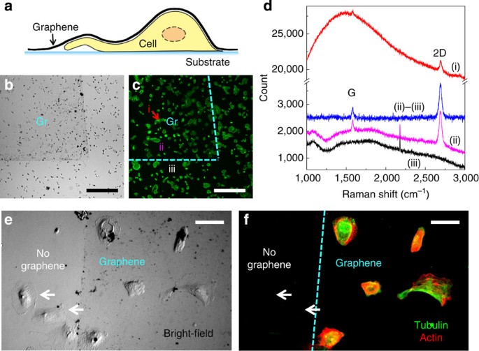
Graphene Enabled Electron Microscopy And Correlated Super Resolution Microscopy Of Wet Cells Nature Communications

Plant Cell Diagram Electron Microscope The Greatest Garden Cell Diagram Animal Cell Structure Plant Cell Diagram

Mrs Mazzuca S Honors Biology Blog Micro Selfies Biology Electron Microscope Scanning Electron Microscope
Cell Organelles Science Learning Hub
Three Dimensional Ultrastructure Of Giant Mitochondria In Human Non Alcoholic Fatty Liver Disease Scientific Reports
Topic 1 2 Ultra Structure Of Cells Amazing World Of Science With Mr Green

Images Of Cilia A Scanning Electron Microscopy Sem Image Of Download Scientific Diagram
What Are The Differences Between A Plant Cell And An Animal Cell

Scanning Electron Microscopy An Overview Sciencedirect Topics

Virus Detection By Transmission Electron Microscopy Still Useful For Diagnosis And A Plus For Biosafety Roingeard 2019 Reviews In Medical Virology Wiley Online Library
![]()
Cytoplasm Definition Function Britannica

Electron Microscopy Atomic Force Microscopy City Of Hope In Southern Ca Electron Microscope Scientific Illustration Microscopy
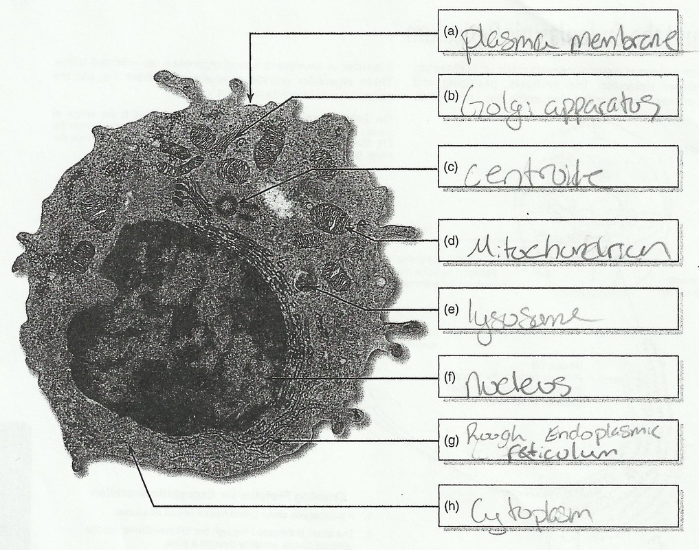
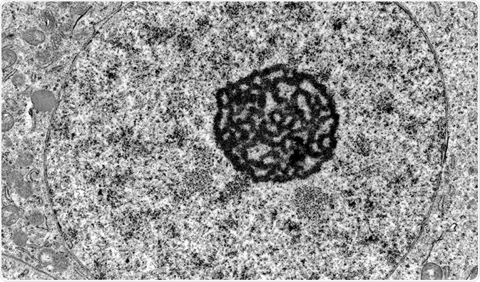
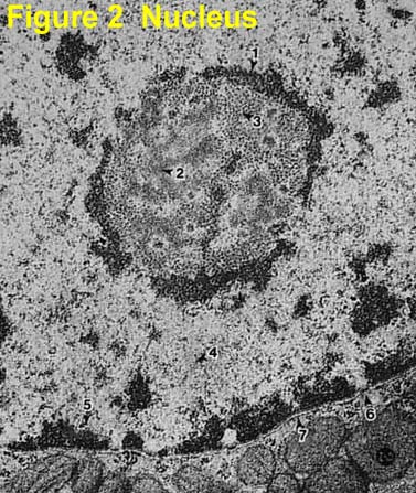
33 Label The Transmission Electron Micrograph Of The Nucleus Label Design Ideas 2020
Scanning Electron Microscopy Of Chromosomes Chapter 9 Scanning Electron Microscopy For The Life Sciences

9 Cell Structure And Function Ideas Cell Structure Structure And Function Cell

1 2 Skill Interpretation Of Electron Micrographs Youtube
Plant Cell Definition Characteristics Facts Britannica

4 3 Eukaryotic Cells Texas Gateway
Ppt Objectives Powerpoint Presentation Free Download Id 6975338

Cytoskeletal Elements
Cellular Organization

How These 26 Things Look Like Under The Microscope With Diagrams
